Ophthalmoscope Ophthalmoscope |
|
|
Diagnostics & Instruments |
|
| |
 |
|
| |
ri-scope®
Ophthalmoscope
|

 |
|
 Direct
ophthalmoscope with XL 3.5 V xenon lamp or HL 2.5 V halogen lamp Direct
ophthalmoscope with XL 3.5 V xenon lamp or HL 2.5 V halogen lamp |
|
For the
examination of narrow and dilated pupils, slit and fluorescence ophthalmoscopy
and the fixation test. |
|
Changes at the front of the eye and to the ocular
fundus become visible |
|
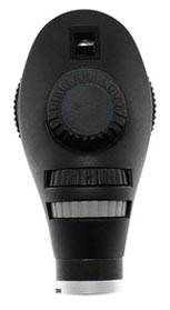
Bayonet fitting
for fast and secure attachment to the handle |
|
Glass-fibre
reinforced casing, particularly sturdy, light-weight and durable |
|
Simple exchange
of the lamp at the base of the instrument head |
|
Focussing wheel
with corrective lenses from 0 to +40 and 0 to –35 dioptres |
|
Completely
shadow-free image thanks to optimum guiding of light beam. Smallest possible
angle between |
|
observational and illuminating
light beams |
|
Illuminated
dioptre display |
|
Illuminated
display of aperture setting |
|
Integrated
eyeglass protector |
|
Easy-to-operate
aperture hand-wheel |
|
Six different
apertures for general and specialist examinations: |
|
Small circle
to reduce reflexes in small pupils |
|
Fixation star
for determining central or eccentric fixation |
|
Semi-circle to
reduce reflexes in small pupils |
|
Grid for
topographical determination of changes in the retina |
|
Large circle
for normal examination of the ocular fundus |
|
Slit for
differential determination, for example, in the case of injuries or tumours |
|
|
Three different
filters: |
|
Red open
filter with contrast-enhancing effect for the exact assessment of fine vascular
changes, |
|
for example,
retinal haemorrhages |
|
Blue filter
for better recognition of vascular anomalies or bleeding, and for fluorescence
ophthalmology |
|
Polarisation
filter for the exact assessment of tissue colour and to avoid corneal
reflections |
|
Each filter can
be used with every aperture. Providing up to 24 examination options |
|
Focussing wheel
to adjust the sharpness of the fixation star image for patients with poor
eyesight |
|
|
|
|
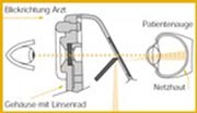
Reflexes from the cornea and
iris are avoided by separating the |
|
observational and
illuminating light beams (Gullstrand’s principle). |
|
The illuminated retinal area
is completely visible – also in the case |
|
of contracted pupils. Optimum
conditions are thus created for |
|
ophthalmological
examinations. |
|
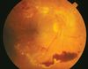
Pathological ocular fundus |
|
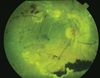
Ocular fundus with red open
filter |
|
|
|
Pathological changes to the ocular fundus can be diagnosed more precisely thanks
to the |
|
numerous examination options |
|
|
|
|
|
|
|
|
|